What happens in the hoof during laminitis?
The hoof corium is a highly vascularized connective tissue between the coffin bone and the hoof capsule. It consists of about 550–600 primary laminae per hoof, each with 100–200 secondary laminae. That adds up to tens of thousands of connection points that anchor the coffin bone in the hoof capsule like Velcro and carry the horse's entire weight [4]. In laminitis, the corium swells severely, which disrupts blood circulation. Because the swelling cannot expand under the rigid hoof capsule, painful pressure builds up. With the lack of blood flow, the corium no longer receives nutrients, the keratinization process stops partially or completely, and the connection between the coffin bone and the hoof capsule breaks down [4].
The coffin bone, no longer securely anchored, can sink downward or rotate around its own axis under the horse's weight and the pull of the deep digital flexor tendon [5]. These mechanical changes are what make laminitis so dangerous, and so painful.
Causes: why does laminitis develop?
The most common cause of laminitis is insulin resistance, the central feature of equine metabolic syndrome (EMS) [3]. Overweight and obesity are the main risk factors for EMS [1]. Horses with EMS often show local fat deposits on the crest, shoulders and tailhead, even when they are not generally overweight. Insulin resistance leads to chronically elevated insulin levels in the blood, and this very hyperinsulinemia is considered the direct trigger of laminitis [2].
Other causes include equine Cushing's disease (PPID), excessive feed intake (especially of carbohydrates such as fructans in spring and fall grass), toxins in the blood during systemic illnesses such as colic, diarrhea or retained placenta, mechanical overload (supporting-limb laminitis) and poisoning, for example from black walnut shavings used as bedding or acorns in large amounts [5].
Hardy breed types such as ponies, Shetland ponies, Icelandic horses and Fjord horses are especially susceptible, as are breeds genetically prone to insulin dysregulation such as the Morgan Horse or certain Quarter Horses [6]. These horses and ponies are often described as “easy keepers” or “good doers.” These are genetically determined traits that offered survival advantages in the wild but can be their undoing in modern horse keeping.
Early detection: warning signs you should know
Many owners do not recognize incipient laminitis in time. In a British study, 45.2% of owners did not recognize the condition in their horse early [7]. The most important early signs:
Bounding digital pulse: A stronger or “pulsating” digital pulse in both front legs is an important early warning sign. In healthy horses, the pulse in the digital arteries is often barely palpable. If it becomes strong and clearly noticeable, it points to inflammation in the hoof.
How to feel the pulse correctly:
- Crouch down beside your horse's front leg.
- Feel with your index and middle fingers (never with your thumb, or you will feel your own pulse!) along the back of the fetlock joint, where the digital artery runs in a groove between the flexor tendon and the suspensory ligament.
- Press gently and stroke sideways until you feel a cord-like bundle of vessels under your fingers.
- A subtle pulse that is hard to find is normal. A strongly throbbing, “pulsating” or “bounding” pulse that you can feel easily is alarming, especially if it appears in both front legs at the same time.
Other early warning signs:
- Warm to hot hooves, especially noticeable at the coronary band
- Short, stilted gait, especially on hard ground
- Horse frequently shifts weight from one leg to the other (“weight shifting”)
- Reluctance to turn or walk downhill
- Palpable or visible rings on the hoof wall (with recurring episodes)
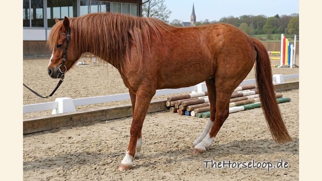
In acute laminitis, symptoms worsen dramatically: the horse shows the typical sawhorse stance, with the front legs placed far forward and the hind legs pushed far under the body to take weight off the painful toes. It lands on the heels (heel-first landing), moves only reluctantly or not at all, shows marked lameness and lies down a lot [5].
Diagnosis and prognosis
Diagnosis is based on a clinical exam, hoof tester examination and X-rays of all affected hooves. Radiographs show whether and how far the coffin bone has already rotated or sunk, which is decisive for the prognosis [5]. The angle between the front edge of the coffin bone and the dorsal wall of the hoof capsule is measured. The larger the deviation, the more severe the laminitis episode and the more guarded the prognosis [5].

The venogram: visualizing the blood supply in the hoof
While X-rays show the bony situation, the venogram (digital venography) provides information about the blood flow to the hoof corium and with it the vitality of the tissue that holds the hoof together [5]. For the exam, a contrast agent containing iodine is injected into the digital vein at the fetlock joint while a tourniquet cuff blocks backflow. The X-ray taken afterward makes the vascular network in the hoof visible.
A healthy hoof shows even, dense contrast filling all the way to the tip of the hoof. In laminitis, by contrast, typical filling defects appear: areas where the blood supply is interrupted, exactly where the hoof corium has been damaged [5]. The larger these defects, the more severe the damage. The venogram helps the vet assess the actual condition of the laminae, give a more precise prognosis and adapt hoof trimming specifically [5]. It is especially valuable in the chronic phase, when a decision has to be made about the further course of therapy.
Prognosis: realistic, but with room to act
Horses with a quick diagnosis and mild coffin bone rotation have good chances of recovery and can return to use as sport or pleasure horses after successful therapy [5]. The earlier therapy is started, the better the chances of a successful recovery [5].
The flip side: in a British cohort study, the consequences of laminitis were one of the most common reasons for euthanasia among the participating horses [6]. The likelihood of a severe course depends, among other things, on when the diagnosis is made, how severe the coffin bone displacement is and how the horse has been used before. These figures underline how important early detection and consistent prevention are. The earlier you watch for warning signs such as increased pulsation and a stiff gait, the better your horse's outlook.
Treatment: what to do in an acute case?
Laminitis is an absolute veterinary emergency. Contact your vet right away if you suspect it. Until the vet arrives:
- Put your horse in a stall with deep bedding (at least 20 cm of peat, sand or shavings) so the hooves sink in and the frog is supported [4].
- Cool the hooves (water hose, ice pack). Cooling eases pain and inflammation.
- Do not feed concentrate, bread or treats. Feed only low-sugar hay in small amounts.
Acute veterinary therapy includes pain medication (nonsteroidal anti-inflammatories such as flunixin meglumine, phenylbutazone), measures to improve circulation, therapeutic hoof care (e.g., with frog pads, plastic wedges or special laminitis shoes) and treatment of the underlying disease, in particular management of insulin resistance and excess weight [5].
Prevention: how to protect your horse
Not every case of laminitis can be prevented, but you can lower the risk considerably:
Weight management: Keep your horse in good body condition. You should be able to feel the ribs but not see them. Excess weight is the biggest risk factor.
Controlled grazing times (in German): Limit turnout in spring and fall, when the fructan content in the grass is especially high. Use a grazing muzzle for at-risk horses.
Low-sugar feeding: Feed a hay-based diet and, if needed, analyze the sugar content of your hay (in German). Soaking can lower the WSC content, but how much depends heavily on the hay: some batches stay above the 10% DM limit even after hours. If in doubt, have it analyzed before and after soaking. A rule of thumb like “60 minutes is enough” is too imprecise. How far the reduction needs to go, when it is needed at all and which feeds are suitable is covered in the article Low-carb horse feeding: what NSC reduction means and when it is needed.
Regular hoof care: Good, physiological hoof trimming by a competent farrier or hoof trimmer every 6–8 weeks.
Monitoring horses at risk: For ponies, easy-keeper breeds and overweight horses, have blood tests for insulin resistance and EMS done regularly.
Exercise: An appropriate training program improves insulin regulation and lowers the risk [3].
Supportive feeding: nutrients for healthy hooves
Good hoof quality can lessen the risk and consequences of chronic laminitis. Several nutrients are well established for horn formation and metabolic stability:
Biotin: The best-known active ingredient for hoof horn quality. Studies show that biotin improves hoof horn formation in horses with poor horn quality. Typical dosage: 15–30 mg per day for at least 6–9 months. Biotin only takes effect over the long term.
Zinc and copper: These trace minerals are essential for keratin formation and the stability of horn structures. An imbalance, often caused by an excess of iron in the base forage, can worsen horn quality. Aim for a balanced ratio (zinc to copper about 3–4 : 1).
Magnesium: In horses with insulin resistance, magnesium supplementation can support insulin regulation. Magnesium is involved in more than 300 metabolic processes, including glucose metabolism. Recommended form: magnesium oxide or organically bound magnesium.
Omega-3 fatty acids (e.g., from flaxseed): Anti-inflammatory properties that can have a supportive effect in chronic inflammatory processes.
Important: Supplements do not replace basic nutrition. The foundation is always needs-based, low-sugar feeding. Talk to your vet or an equine nutritionist about supplementation for horses with metabolic disease or at risk of laminitis.
Bottom line
Laminitis is a serious, painful condition, but with knowledge, vigilance and quick action you can considerably improve your horse's chances of recovery and quality of life. Learn the early warning signs, check the digital pulse regularly, and watch for warm hooves and changed movement patterns. If you suspect it, don't hesitate: every hour counts. Prevention through weight management, controlled turnout times and regular hoof care remains the best protection against this devastating condition.
Frequently asked questions about laminitis in horses
How does laminitis develop in horses?
In laminitis, the hoof corium between the coffin bone and the hoof capsule swells severely, which disrupts blood circulation [4]. Because the swelling cannot expand under the rigid hoof capsule, painful pressure builds up, and the undersupplied corium loses its anchoring function [4]. As a result, the connection between the coffin bone and the hoof capsule can loosen, so that the coffin bone can sink or rotate under the weight and the pull of the deep digital flexor tendon [5].
Why do horses get laminitis?
The most common cause is insulin resistance, the central feature of equine metabolic syndrome, with excess weight being the biggest risk factor [3][1]. Chronically elevated insulin levels in the blood are considered a direct trigger of laminitis [2]. Other causes include equine Cushing's disease, excessive intake of carbohydrates such as fructans from grass, toxins in conditions such as colic or diarrhea, mechanical overload and poisonings, for example from acorns in larger amounts [5].
Can any horse get laminitis?
In general, laminitis can affect many horses, but hardy breed types such as ponies, Shetland ponies, Icelandic horses and Fjord horses are especially susceptible, as are breeds genetically prone to insulin dysregulation, such as the Morgan Horse or certain Quarter Horses [6]. These horses are often described as “easy keepers” or “good feed converters.” That was an advantage in the wild, but it can become a risk in modern horse keeping. Overweight horses and horses with EMS are at particular risk regardless of breed [1].
How do I recognize laminitis in horses?
An important early warning sign is an increased, clearly palpable pulse in the digital arteries, especially if it appears in both front legs at the same time. Other signs are warm to hot hooves, a cautious, stilted gait on hard ground, frequent shifting of weight from one leg to the other, and reluctance to turn or go downhill. In acute laminitis, your horse shows the typical sawhorse stance with the front legs placed far forward, puts weight on the heels and lies down a lot [5].
What should I do if my horse has laminitis?
Laminitis is an absolute veterinary emergency. If you suspect it, contact your vet right away. Until your vet arrives, stand your horse on deep bedding of at least 20 cm of peat, sand or shavings, so the hooves sink in and the frog is supported [4]. Cool the hooves with a hose or an ice pack, which eases pain and inflammation. Feed only low-sugar hay in small amounts, no concentrate, bread or treats.
Which drugs and painkillers does the vet give for laminitis?
Acute veterinary treatment includes pain medication with nonsteroidal anti-inflammatories such as flunixin meglumine or phenylbutazone [5]. It also includes measures to improve circulation, therapeutic hoof care, for example with frog pads, plastic wedges or special laminitis shoes, and treatment of the underlying disease, especially management of insulin resistance and excess weight [5]. Dosages and the choice of drugs always belong in the hands of your vet.
Is laminitis in horses curable?
Horses diagnosed quickly and with mild coffin bone rotation have a good chance of recovery and, after successful treatment, can return to use as sport or pleasure horses [5]. The earlier treatment starts, the better the chances of a successful recovery [5]. In a British cohort study, however, the consequences of laminitis were one of the most common reasons for euthanasia among the participating horses [6]. This underlines how crucial early detection is. Decisions like that always belong in a conversation with your vet.
Which herbs or homeopathic remedies help with laminitis?
The sources reviewed here contain no evidence of effectiveness for herbs or homeopathic remedies in laminitis, and supplements never replace basic care. What is documented, however, is that biotin can improve hoof horn formation in the long term when hoof horn quality is poor. It does not help in the acute phase. Because laminitis is an emergency, none of this replaces immediate veterinary treatment, and you should discuss supplementation for horses at risk of laminitis with your vet or an equine nutritionist.
What can horses with laminitis eat?
In an acute case, feed only low-sugar hay in small amounts and skip concentrate, bread and treats. The basis is generally a needs-based, low-sugar, hay-based diet, and if needed, the sugar content of the hay can be analyzed. Soaking can lower the WSC content, but how much depends heavily on the hay, so some batches stay above the 10% limit even after hours. If in doubt, have the hay analyzed before and after soaking. A rule of thumb like “60 minutes is enough” is too imprecise.
How much exercise makes sense for a horse with laminitis?
In an acute case, your horse belongs in a stall with deep bedding, so the hooves sink in and the frog is supported [4]. Exercise plays a role mainly in prevention: an appropriate training program improves insulin regulation and reduces the risk [3]. When and how your horse may be exercised again after a flare-up depends on the individual case and should be discussed with your vet.
Sources
- [1] Johnson PJ et al. - Laminitis and the equine metabolic syndrome. Vet Clin North Am Equine Pract 2010. https://pubmed.ncbi.nlm.nih.gov/20699172/
- [2] McGowan CM - Equine metabolic syndrome. In Practice 2015. https://pmc.ncbi.nlm.nih.gov/articles/PMC4552932/
- [3] Durham AE et al. - ECEIM consensus statement on equine metabolic syndrome. J Vet Intern Med 2019. https://pubmed.ncbi.nlm.nih.gov/30724412/
- [4] Pollitt CC. - The anatomy and physiology of the suspensory apparatus of the distal phalanx. Vet Clin North Am Equine Pract. 2010;26(1):29–49. https://pubmed.ncbi.nlm.nih.gov/20381734/
- [5] Belknap JK. - The pharmacologic basis for the treatment of developmental and acute laminitis. Vet Clin North Am Equine Pract. 2010;26(1):115–124. https://pubmed.ncbi.nlm.nih.gov/20381740/
- [6] Pollard D, Wylie CE, Newton JR, Verheyen KLP - Factors associated with euthanasia in horses and ponies enrolled in a laminitis cohort study in Great Britain. Prev Vet Med. 2020. https://pubmed.ncbi.nlm.nih.gov/31751854/
- [7] Pollard D, Wylie CE, Verheyen KLP, Newton JR. - Assessment of horse owners' ability to recognise equine laminitis: A cross-sectional study of 93 veterinary diagnosed cases in Great Britain. Equine Vet J. 2017;49(6):759–766. https://pubmed.ncbi.nlm.nih.gov/28543943/
All medical statements in the text are linked to the corresponding source in this list with inline markers [N]. This article does not replace advice from your vet.


